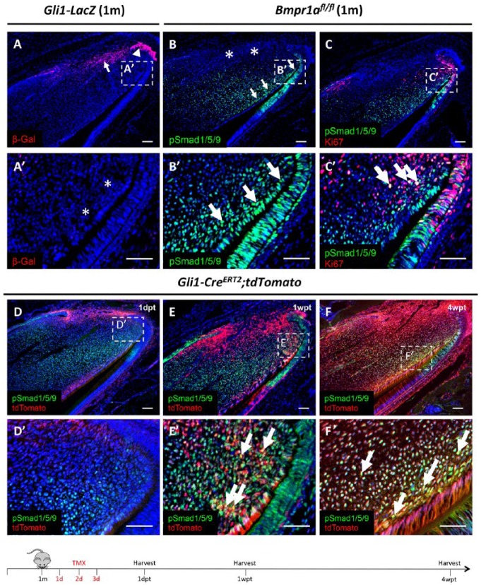
Figure 1.
Activation of bone morphogenetic protein (BMP) signaling in Gli1+ mesenchymal stem cell (MSC)–derived preodontoblasts/odontoblasts in adult mouse incisors. (A) β-Gal immunostaining (red) of sagittal sections of incisors from 1-mo-old (1m) Gli1-LacZ mice. Arrow and arrowhead indicate Gli1+ cells in the proximal end of the incisor epithelium and mesenchyme, respectively. (A′) Higher magnification of boxed region in A shows absence of Gli1 expression in the preodontoblast/odontoblast region indicated by asterisks. (B, B′) pSmad1/5/9 immunostaining (green) of sagittal sections of incisors from 1-mo-old (1m) Bmpr1afl/fl mice. Arrows indicate pSmad1/5/9 signaling in the preodontoblast/odontoblast region, and asterisks indicate absence of pSmad1/5/9 signaling in the Gli1+ MSC region. Boxed area in B is shown magnified in B′. (C) pSmad1/5/9 (green) and Ki-67 (red) double immunostaining of sagittal sections of incisors from 1-mo-old (1m) Bmpr1afl/fl mice. Boxed area in C is shown magnified in C′. Arrows indicate colocalization of Ki-67+ cells and BMP signaling activity (yellow). (D–F′) Lineage tracing of sagittal sections of incisors from 1-mo-old Gli1-CreERT2;tdTomato mice 1 d (1dpt), 1 wk (1wpt), and 4 wk (4wpt) posttamoxifen induction. Red indicates Gli1-derived cells; green indicates BMP signaling activity; yellow indicates colocalization of fluorescent staining (arrows). Boxes in D to F are shown magnified in D′ to F′, respectively. Schematic diagram at the bottom indicates induction protocol. Scale bars, 100 µm.

