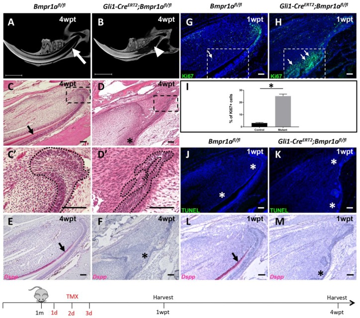
Figure 2.
Loss of bone morphogenetic protein (BMP) signaling in Gli1+ cells arrests incisor growth. (A, B) Micro–computed tomography analysis of incisors from 1-mo-old Bmpr1afl/fl (A) and Gli1-CreERT2;Bmpr1afl/fl (B) mice 4 wk after tamoxifen induction (4wpt). Arrow indicates proximal end of Bmpr1afl/fl incisors and arrowhead indicates lack of proximal end of Gli1-CreERT2;Bmpr1afl/fl incisors. (C, D′) Hematoxylin and eosin staining of sagittal sections of mandibular incisors from 1-mo-old Bmpr1afl/fl (C, C′) and Gli1-CreERT2;Bmpr1afl/fl (D, D′) mice 4 wk after tamoxifen induction (4wpt). Boxes in C and D are magnified in C′ and D′, respectively. Arrow in C indicates dentin formation and asterisk in D indicates absence of dentin formation. Dotted lines in C′ and D′ indicate the cervical loop’s outline. (E, F) Dspp RNAscope in situ hybridization (red) of sagittal sections of mandibular incisors from control (E) and Gli1-CreERT2;Bmpr1afl/fl (F) mice 4 wk after tamoxifen (4wpt) induction at 1 mo of age. Arrow indicates Dspp+ odontoblasts and asterisk indicates absence of Dspp expression. (G, H) Ki-67 immunostaining (green) of sagittal sections of mandibular incisors from 1-mo-old Bmpr1afl/fl (G) and Gli1-CreERT2;Bmpr1afl/fl (H) mice 1 wk after tamoxifen induction (1wpt). Arrows indicate Ki-67 expression in the preodontoblast/odontoblast region (boxed area) of the incisor. (I) Quantitation of Ki-67+ cells in Bmpr1afl/fl (control) and Gli1-CreERT2;Bmpr1afl/fl (mutant) incisor odontogenic corresponding to the boxed areas in G and H, respectively. Quantitation was performed by calculating the percentage of Ki-67+ cells per section (n = 4). *P < 0.05. (J, K) Terminal deoxynucleotidyltransferase-mediated dUTP nick end labeling (TUNEL) staining (green) of sagittal sections of mandibular incisors from 1-mo-old Bmpr1αfl/fl control (J) and Gli1-CreERT2;Bmpr1afl/fl (K) mice 1 wk after tamoxifen induction (1wpt). Asterisks in J and K indicate absence of apoptotic activities in the entire proximal ends of incisors. (L, M) Dspp RNAscope in situ hybridization (red) of sagittal sections of mandibular incisors from 1-mo-old Bmpr1αfl/fl control (L) and Gli1-CreER;Bmpr1αfl/fl (M) mice 1 wk after tamoxifen induction (1wpt). Arrow in L indicates Dspp+ odontoblasts in the control odontogenic region, and asterisk in M indicates absence of signaling in the Gli1-CreERT2;Bmpr1αfl/fl odontogenic region. Schematic diagram at the bottom indicates induction protocol. Scale bars (A, B) 150μm; (C–H, J–M), 100 µm.

