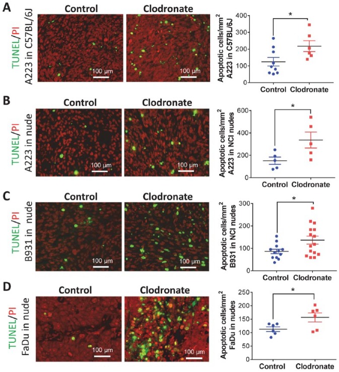
Figure 3.
Increased apoptosis in squamous cell carcinomas (SCCs) from clodronate-treated mice. Apoptotic cells were identified by TUNEL staining and nuclei by propidium iodide (PI) staining, quantified in each SCC model, and analyzed by Student’s t test. (A) A223 tumors grown in C57BL/6J mice. (B) A223 tumors grown in athymic nude mice. (C) B931 tumors grown in athymic nude mice. (D) FaDu tumors grown in athymic nude mice. Values are presented as mean ± SEM. *P < 0.05.

