Figure 5.
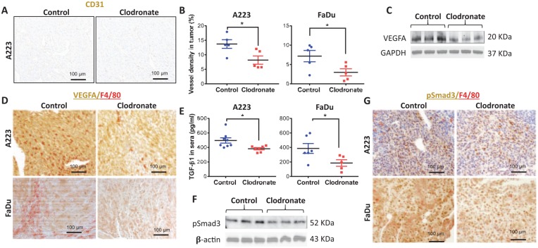
Macrophage-depleted tumors have decreased blood vessel density and angiogenic factors. (A) Blood vessels in A223 tumors in C57BL/6J mice were identified by immunohistochemistry staining for endothelium marker CD31. (B) Vessel lumen size was quantified as a percentage of tumor area for A223 and FaDu tumors. (C) Levels of VEGFA and GAPDH in A223 tumor lysates were determined by Western blot analysis. (D) Immunohistochemistry staining of VEGFA and F4/80 in A223 tumors grown in C57BL/6J mice or FaDu tumors grown in athymic nude mice. (E) An ELISA kit was used to detect TGFβ1 levels in the sera of A223 tumor-bearing C57BL/6J mice or FaDu tumor-bearing athymic nude mice treated with control or clodronate liposomes. (F) Levels of pSmad3 and β-actin in A223 tumor lysates were determined by Western blot analysis. (G) Immunohistochemistry staining of pSmad3 and F4/80 in A223 tumors grown in C57BL/6J mice and FaDu tumors grown in athymic nude mice. Statistics by Student’s t test; *P < 0.05. (B, E) Values are presented as mean ± SEM.

