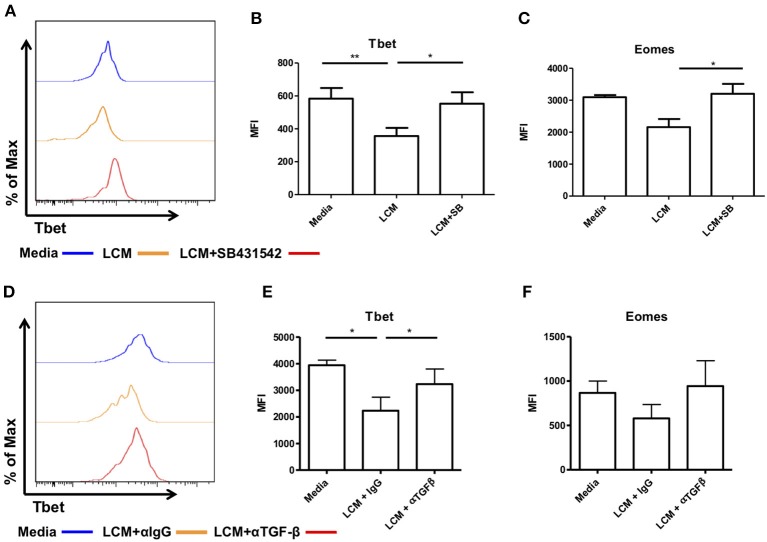
Figure 6.
Blocking TGF-β inhibits the ability of liver conditioned media to suppress Tbet and Eomes expression. CD56bright Eomeshi Tbetlo NK cells FACS isolated from liver perfusate (LP) were cultured for 7 days, supplemented with 10% v/v liver conditioned media (LCM) with and without SMAD inhibitor SB431542 and acquired on a FACS Fortessa. (A) Representative histogram of Tbet expression from cells at day 7 untreated (blue line), 10% LCM (orange line) or LCM & SB431542 (red line). (B) MFI of Tbet in CD56bright NK cells from LP at day 7 with LCM or LCM & SB431542. (C) MFI of Eomes in CD56bright NK cells from LP at day 7 with LCM or LCM & SB431542. NK cells isolated from peripheral blood (PB) were cultured for 7 days, supplemented with 10% v/v LCM with 5 μg/mL anti-TGF-β1 blocking antibody or IgG isotype control and acquired on a FACS Canto II. (D) Representative histogram of Tbet expression in CD56bright NK cells from peripheral blood at day 7 with LCM or LCM & TGF-β1 blocking antibody. (E) MFI of Tbet in CD56bright NK cells from peripheral blood at day 7 with LCM or LCM & TGF-β1 blocking antibody. (F) MFI of Eomes in CD56bright NK cells from peripheral blood at day 7 with LCM or LCM & TGF-β1 blocking antibody. Data presented as mean ± SEM. Data was analyzed using Friedman test, with Dunn's multiple comparison test or repeated measures one-way ANOVA (n = 3–8, *p < 0.05 and **p < 0.01).

