
Figure 1.
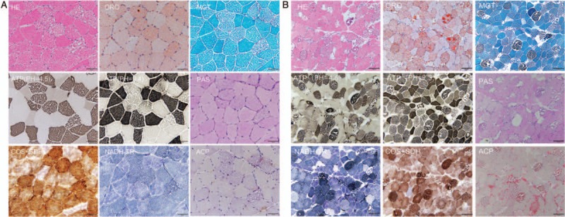
Muscle pathology of the Patient 1 (A) and Patient 2 (B). (A) HE staining showed a numerous of large vacuoles deposited in muscle fibers. ORO staining revealed that vacuoles were stained by ORO dye, indicating the accumulation of lipid droplets. NADH-TR staining revealed a slightly abnormal structure in type II fibers. Cyclo-oxygenase and SDH recombination staining showed enzymatic deficiency in some fibers. Scale bar = 50 μm. (B) The characteristic features of Patient 2 were highly similar to those of Patient 1. However, there were many scattered necrotic fibers in the type I myofibers. MGT staining displayed many dark blue necrotic fibers. Scale bar = 100 μm. HE: Hematoxylin-eosin; MGT: Modified gomori trichrome; NADH-TR: Nicotinamide adenine dinucleotide-tetrazolium reductase; ORO: Oil red O; SDH: Succinate dehydrogenase.