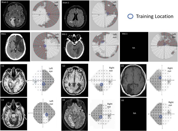
Figure 3.
Neuroradiological images and visual perimetries of CB patients. All patients sustained damage of early visual areas or the optic radiations, resulting in homonymous visual field defects as shown by the visual field perimetries, next to each brain image. Within the perimetry images (patients in top two rows: Sham1, Sham2, RNS1, RNS2, and RNS3): red marks and shading areas indicate the patients' blind field. Bottom two rows: Humphrey visual field maps for each of the unstimulated patient (U1–6), with superimposed shading indicating the blind field and numbers indicating the luminance detection sensitivity in the given position expressed in decibels. For all patients, the blue circles indicate the training location and size (for details, see Global direction discrimination testing and training in patients, in Materials and Methods). Radiological images were not available for patients RNS3 and U6.

