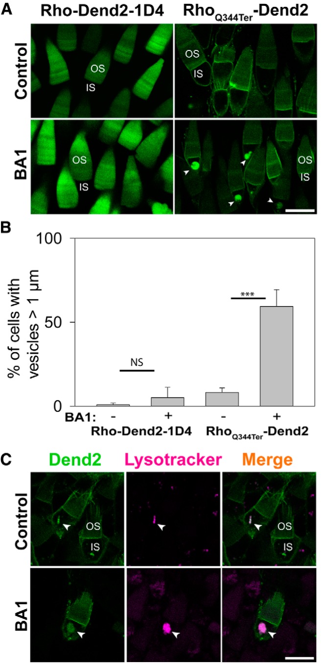
Figure 1.

Class I mutant rhodopsin accumulates in lysosomes after inhibition of lysosome-mediated degradation. A, Confocal imaging of live Xenopus laevis rod photoreceptors expressing either Rho-Dend2-1D4 (left) or RhoQ344ter-Dend2 (right). OS and IS structures are labeled in a representative rod cell of each image. Before the imaging, the animals were treated with DMSO (Control) or 100 nm BA1 (BA1) for 24 h. In control retinas, neither Rho-Dend2-1D4 nor RhoQ344ter-Dend2 accumulated significantly in vesicular structures of the ISs. After BA1 treatment, RhoQ344ter-Dend2 accumulated in vesicular structures (arrowheads) in the ISs, but Rho-Dend2-1D4 did not. B, In retinas expressing either Rho-Dend2-1D4 or RhoQ344ter-Dend2, we assessed the percentage of rod photoreceptor cells that exhibited vesicles >1 μm in diameter. BA1 treatment of photoreceptors expressing Rho-Dend2-1D4 increased this percentage from 0.9 ± 1.0% (untreated) to 5.1 ± 6.2% (NS, p = 0.23). BA1 treatment of photoreceptors expressing RhoQ344ter-Dend2 increased this percentage from 8.2 ± 2.7% to 59.4 ± 10.0% (p < 0.001), indicating that RhoQ344ter-Dend2, but not Rho-Dend2-1D4, accumulates upon BA1 treatment. C, Confocal imaging of live Xenopus laevis rod photoreceptors expressing RhoQ344ter-Dend2 and labeled with LysoTracker Red dye. LysoTracker Red signal colocalized with RhoQ344ter-Dend2 in untreated cells (Control, arrowheads), and the amount of RhoQ344ter-Dend2 colocalized with LysoTracker Red increased after pretreatment with BA1 (arrowheads). Scale bars, 10 μm. ***p < 0.001, NS, not significant.
