Figure 2.
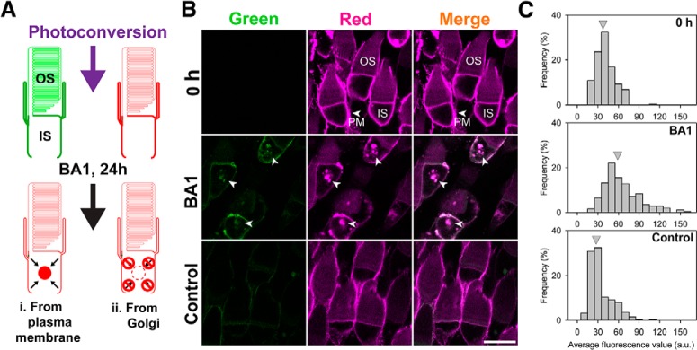
Mislocalized rhodopsin is trafficked from the plasma membrane to intracellular lysosomes. A, Experimental design to determine the origin of proteins accumulating in the ISs of RhoQ344ter-Dend2 cells. Before photoconversion, the majority of RhoQ344ter-Dend2 is localized to the IS PM. After photoconversion and BA1 treatment (24 h), red protein should accumulate if RhoQ344ter-Dend2 is being trafficked from the PM to the IS lysosomes (Model i). If RhoQ344ter-Dend2 is not internalized, then red RhoQ344ter-Dend2 will not accumulate significantly in the IS lysosomes (Model ii). B, Immediately following photoconversion, rods expressing RhoQ344ter-Dend2 were imaged (0 h). Photoconversion was efficient as confirmed by the loss of green fluorescence (0 h, green). The majority of red RhoQ344ter-Dend2 was observed on the IS PM (0 h, red, PM, arrowhead). After 24 h of BA1 treatment, RhoQ344ter-Dend2 accumulated intracellularly in the ISs of rods (BA1, arrowheads). A nominal amount of green RhoQ344ter-Dend2 was observed after 24 h (BA1 and Control, Green). This observation indicates that new RhoQ344ter-Dend2 was synthesized in these retinas. C, Average red fluorescence intensities (a.u./area) were measured for intracellular regions of ISs in individual rods. The histograms indicate frequency of cells (y-axis) for each fluorescence intensity range (x-axis). Gray arrowheads indicate the average fluorescence intensity of each condition (0 h, BA1, or Control). Red fluorescence intensity values in the ISs were 36.7 ± 13.9 a.u. for time = 0 h condition, 29.4 ± 16.8 a.u. for 24 h DMSO-treated Control group, and 60.2 ± 16.8 a.u. for the BA1-treated group. Animals were aged 9 DPF upon photoconversion (0 h) and were killed and imaged at 10 DPF (BA1 and Control), with a total of 250 cells for each condition measured from n = 5 animals. Scale bar, 10 μm.

