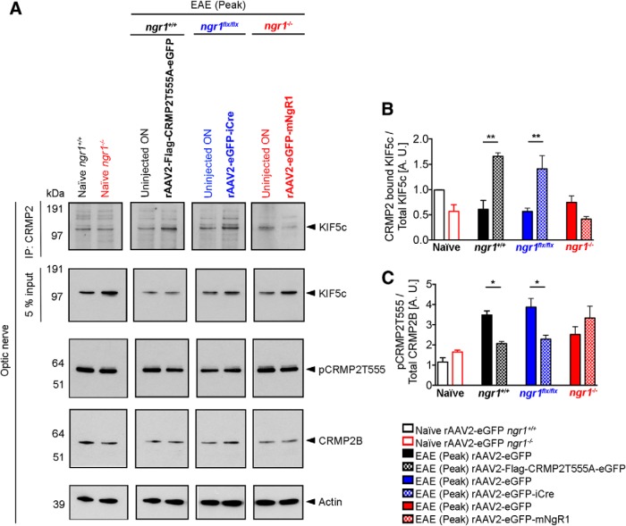
Figure 4.
Axon-specific deletion of ngr1 maintains the protein interactions between CRMP2 and KIF5c in the optic nerve during EAE. A, First row, Immunoprecipitation of CRMP2 of optic nerve lysates from naive ngr1+/+ and ngr1−/−; EAE-induced optic nerve lysates (at peak stage of disease) from either: ngr1+/+ transduced with rAAV2-CRMP2T555A-eGFP, ngr1flx/flx transduced with rAAV2-eGFP-iCre, ngr1−/− mice transduced with rAAV2-eGFP-mNgR1 and their left (contralateral) uninjected optic nerve controls. The membranes were then re-probed using anti-KIF5c antibody. Second row, A pre-immunoprecipitation 5% input of total protein. Third row, Western immunoblot detection of the phosphorylation of CRMP2 at the threonine 555 site (pCRMP2-T555), (fourth row) total CRMP2, and (fifth row) actin loading control. B, Densitometric quantification of total KIF5c and CRMP2 bound KIF5c (after immunoprecipitation) from optic nerve lysates. C, Densitometric quantification of level of phosphorylation of CRMP2B at T555 over total CRMP2B (n = 3, mean ± SEM, one-way ANOVA with post Tukey's test; *p < 0.05, **p < 0.01).

