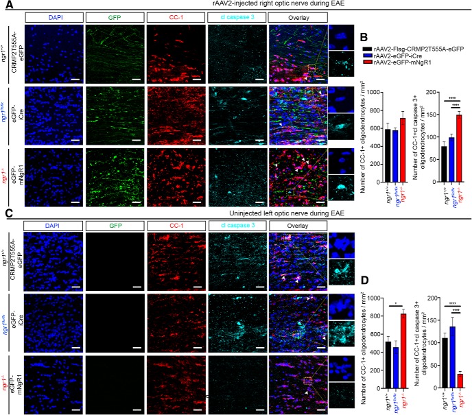
Figure 6.
Axon-specific deletion of ngr1 protects oligodendrocytes during EAE. Representative images of apoptotic CC-1-positive mature oligodendrocytes, expressing cleaved caspase-3 in (A) rAAV2-injected right optic nerves, and (C) rAAV2-uninjected left optic nerves. Scale bar, 50 μm. High-power images show anti-activated caspase 3 staining and the presence of pyknotic nuclei (from yellow dotted box). Number of CC-1-positive mature oligodendrocytes and cleaved caspase-3-immunopositive oligodendrocytes per square millimeter of (B) right and (D) left optic nerves at the peak stage of EAE (n = 4, mean ± SEM, one-way ANOVA with post Tukey's test; *p < 0.05, ****p < 0.0001). Arrowheads indicate apoptotic mature oligodendrocytes. For the number of CC-1+ mature oligodendrocytes in optic nerves of naive ngr1+/+ and ngr1−/− mice, see Figure 6-1.

