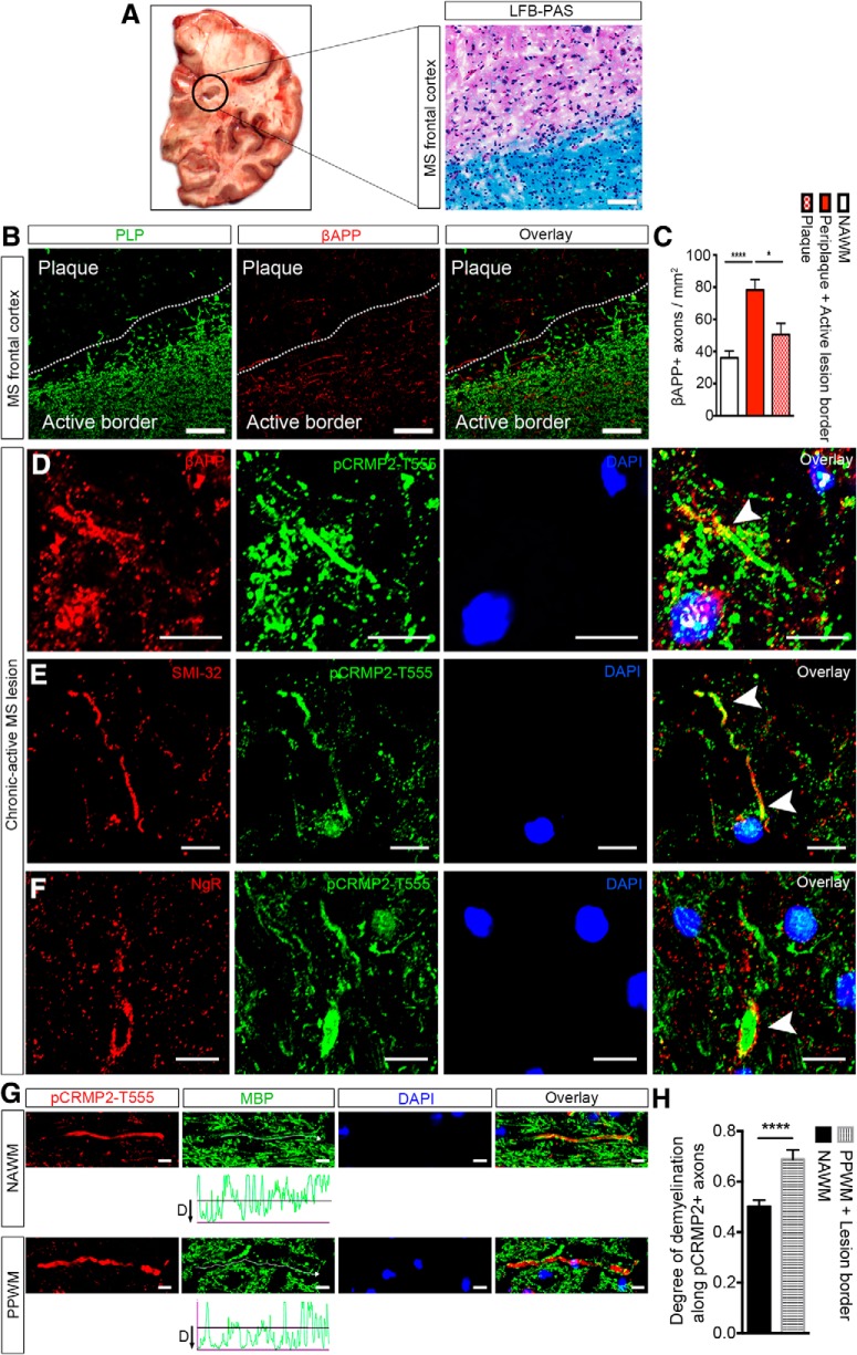
Figure 7.
pCRMP2-T555 localization in degenerating neuronal somata and axons in chronic-active lesions of MS brains. A, LFB-PAS immunohistochemistry showing representative chronic demyelinated lesion from secondary progressive MS frontal cortex. B, Immunostaining against anti-proteolipid protein (PLP; green) and anti-βAPP (red) on serial section showing significant numbers of βAPP-positive axons in a representative image of an active demyelinating lesion (active border) and a few βAPP-positive axons in an inactive demyelinated plaque. Scale bar, 100 μm. C, βAPP expression in MS lesions of different demyelinating activity [NAWM, periplaque along with the active lesion border (active demyelinating lesion), plaque (inactive demyelinated lesion); n = 4, mean ± SEM, one-way ANOVA with post Tukey's test; *p < 0.05, ****p < 0.0001]. Expression of pCRMP2-T555 alongside common neurodegenerative markers: (D) pCRMP2-T555 expression in the distal segment of an βAPP-positive degenerating axon, and (E) colocalization with non-phosphorylated SMI-32-positive neurofilament. F, Degenerative ovoid formation and swelling in pCRMP2-T555-positive axons in human deep cortical white matter. Arrowheads indicate extracellular localization of NgR and downstream intracellular phosphorylation of CRMP2 in a degenerative axon. G, Immunohistochemistry showing pCRMP2-T555 (red), MBP (green) in NAWM and PPWM. H, The degree of demyelination around pCRMP2-T555-positive axons is significantly higher within active demyelination lesions compared with NAWM (n = 4, mean ± SEM, Student's t test; ****p < 0.0001). Scale bar, 10 μm.

