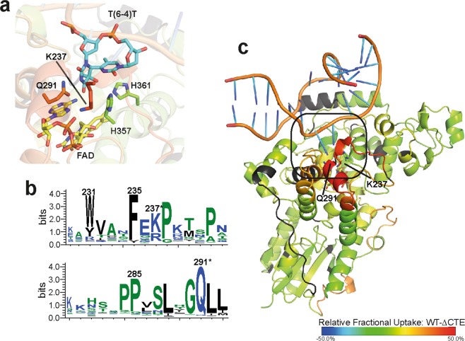
Figure 3.
Closer view on the DNA binding site of CraCRY. For that the relative uptake of the FADOX state at 10 s was mapped onto the CraCRY∆CTE-DNA structure (PDB entry: 6FN0). (a) Here, the (6-4) damaged DNA (blue) is bound to the enzyme and the two catalytic histidines (green) are shown. (b) Q291 and K237 play a crucial role in DNA binding and are highly conserved (marked with *) as shown in the WebLogo that was generated from a multiple sequence alignment of animal-like cryptochromes8. Both residues are located on a stretch with high deuterium uptake in the WT. (c) The overall binding mode of the dsDNA is expected to be unaffected between CraCRY-WT and CraCRY∆CTE when taking the HDX analysis results without DNA into account.

