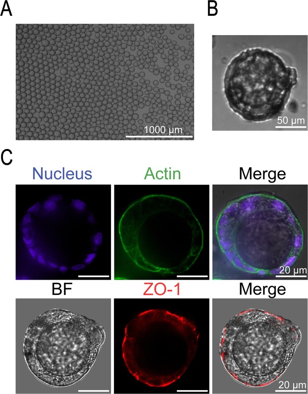
Figure 6.
Reversal of apical-basal polarity using Matrigel beads. (A) Representative image of Matrigel beads in oil (30–60 μm diameter). (B) Brightfield image showing Matrigel bead surrounded by cells in an agarose microwell (10X). (C) Fluorescent images (40X) showing a continuous monolayer of cells surrounding a Matrigel bead shown by the DAPI stain (blue) and reversal of polarity with actin (green) and tight junction protein ZO-1 (red) expressed on the outer membrane away from the Matrigel bead.

