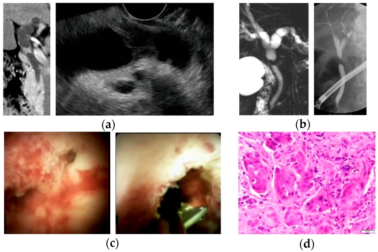
Figure 2.
A case of bile duct stricture diagnosed as perihilar cholangiocarcinoma with peroral cholangioscopy (POCS)-guided forceps biopsy. (a) Computed tomography scan and endoscopic ultrasonography showed an irregular nodule in the perihilar bile duct; (b) magnetic resonance cholangiopancreatography and endoscopic retrograde cholangiography revealed irregular stenosis in the perihilar bile duct; (c) POCS showed the irregular papillary mucosa that existed from the bifurcation of the cystic duct to the perihilar bile duct. POCS-guided forceps biopsy was performed for the biliary stricture in the perihilar bile duct; and (d) hematoxylin and eosin staining revealed adenocarcinoma in specimens obtained from the biliary stricture.

