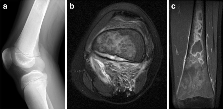
Fig. 10.
Bone infarct and subperiosteal hemorrhage mimicking osteomyelitis in Gaucher disease. A 13-year-old male presented with atraumatic knee pain and swelling. Initial lateral knee radiograph (a) showed subtle periosteal reaction along the posterior metadiaphysis of the distal femur and ill-defined intramedullary sclerosis within the femoral shaft. Follow-up MRI demonstrated intramedullary marrow edema and focal subperiosteal fluid with surrounding inflammatory changes in the popliteal fossa thought to be related to subperiosteal hemorrhage on axial T2-weighted image (b). Coronal post-contrast T1 fat-saturated image (c) demonstrated peripheral serpentine enhancement of areas of bone infarction within the distal femur. The patient was subsequently diagnosed with Gaucher disease after initial workup for hematopoietic malignancy

