Fig. 2.
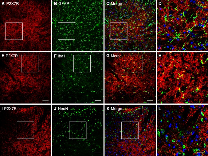
Cellular localization of P2X7R immunoreactivity in the rat spinal dorsal horn. A–L Cell-type-specific immunolabeling of P2X7Rs in the ipsilateral dorsal horn at 4 h after intraplantar BmK I administration. (A, E, I) P2X7R-positive staining revealed no P2X7R co-localization with either the astrocytic marker GFAP or the neuronal marker NeuN (B, C, J, K). F–G Double immunofluorescence in the superficial dorsal horn showed that P2X7Rs co-localized with the microglial marker Iba-1. White open squares in C, G, K indicated the corresponding magnified images (D, H, L) in the confocal images. Scale bars, 50 μm in A–C, E–G, I–K; 20 μm in D, H, L.

