Fig. 3.
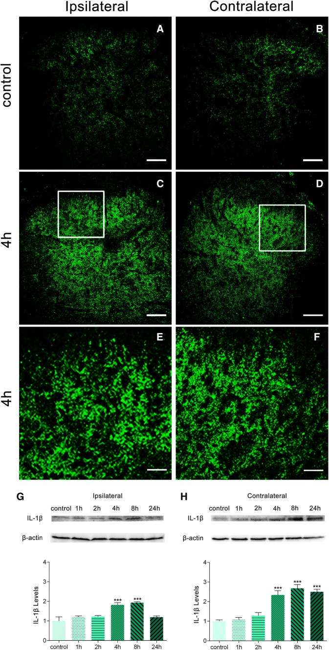
Effects of BmK I on the release of IL-1β in the dorsal horn. Immunoreactivity of IL-1β in the rat spinal cord in the presence of BmK I. A–F Compared with the control group (A, B), bilateral IL-1β immunoreactivity in the dorsal horn increased significantly in BmK I-treated rats (C–F). White squares in C and D indicate the magnified images in E and F. Scale bars, 100 μm in A–D; 50 μm in E, F. G–H Representative Western blots of IL-1β and β-actin in the ipsilateral (G) and contralateral (H) spinal cord; histograms show the mean levels with respect to each control group at different time points after intraplantar BmK I injection. The data are presented as mean ± SEM (n = 3; ***P < 0.001 compared with control, one-way ANOVA followed by Dunnett’s post hoc test).

