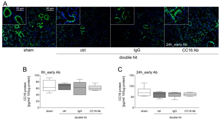
Figure 2.
(A) CC16 (green) and nuclei (blue, 4′,6-diamidino-2-phenylindole, DAPI) staining 24 hours after CLP and early interventions with antibodies shows intrabronchial high concentrations of CC16 positive cells. In double-hit groups, a prominent loss of CC16 into the interstitium as well as a loss of the pulmonary integrity is shown. (B) Pulmonary CC16 levels are shown in animals with early interventions with either CC16 antibody (Ab) or IgG control (IgG) antibody, at 6 or 24 h post-CLP (C) (n = 8).

