Figure 1.
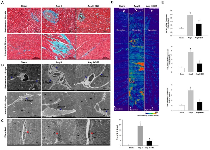
Simvastatin suppresses in vivo angiotensin (Ang) II-mediated collagen deposition, fibrosis, and collagen-associated protein expression. Male Sprague-Dawley rats were treated with (Ang II, 1 mg/kg/day) or Ang II + simvastatin (SIM, oral, 10 mg/kg) for 28 days. Perivascular and interstitial fibrosis ultrastructures (blue color) were determined using Masson’s trichrome staining. (A) The collagen fibers are shown in the representative images (blue) (n = 6). Perivascular and interstitial collagen fiber expression (blue arrows) (B) and quantification by transmission electron microscopy (TEM) analysis are shown (Supplemental Figure S1A,B). The cardiac fibroblast (CF) ultrastructures (red arrows) were determined using TEM analysis (C) (n = 6) (mf, myofibril; m, mitochondrial; CF, cardiac fibroblast). Cardiac collagen deposition and arrangement from endocardium to epicardium were identified and quantified using second-harmonic generation (SHG) microscopy (D) (n = 3). * p < 0.05 vs. the control from three independent experiments. The fibrotic associated genes: ACTA2, COL1A1, and LOXL2 expression were analyzed using real-time PCR and normalized by β–actin (E). For all comparisons, * p < 0.05 vs. sham.

