Figure 3.
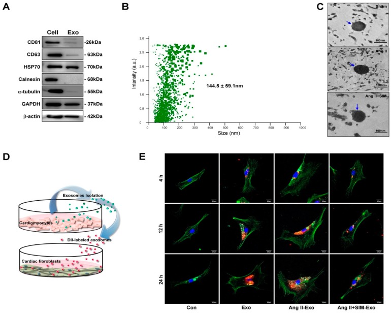
HCM-derived exosomes are transported into human cardiac fibroblasts (HCF) in vitro. Exosomal-specific marker expression from HCM-isolated exosomes was measured using Western blot analysis (A). HCM-isolated exosome size was demonstrated by NanoSight NS300 (B) and TEM analysis (C). To identify uptake of HCM-derived exosomes by HCF, exosomes were isolated from HCM-conditioned medium, after treatment with Ang II, or Ang II + SIM for 24 h. HCM exosomes were labeled with the DiI dye (5 μg/mL, red florescence) for 1 h, and incubated with HCF cells for various times (4, 12, and 24 h) (D). HCF cells were non-treated (control), or treated with Eox, Ang II-Exo and Ang II-SIM-Exo for various times (4, 12, and 24 h), then stained for intracellular cytoskeleton F-actin (green florescence; nucleus, blue florescence), and analyzed by confocal microscopy (E). Scale bars: 20 μm. Each image is representative of three independent experiments.

