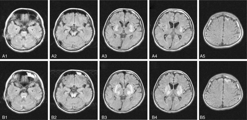
Figure 2.

Brain magnetic resonance imaging (MRI) in a patient with Wilson disease (WD) before and after a traumatic event. These images were from a 25 years old female patient who had been affected with WD with mixed hepatic and neurological presentations for 2 years. Before trauma (A1-A5), the neurological symptoms were slurred speech and drooling, and these symptoms became more severe to be both hand and head tremors and clumsy movement apart from the initial symptoms of slurred speech and drooling 1 hour after falling down with head impacted on ground while walking (B1-B5). Three days before trauma (A1-A5), high signals in bilateral Pons (A1), midbrain (A2), putamen (A3, A4), and left frontal lobe (A5) on flair-weighted MRI. One hour after trauma (B1-B5), apart from the high signal in Pons (B1), the areas of high signals in midbrain(B2), putamen (B3, B4), and left frontal lobe (B5) on flair weighted MRI became wider than those before trauma, and high signals in bilateral thalamus (B3 and B4) and caudate nucleus (B3) were also noticed after traumatic event.
