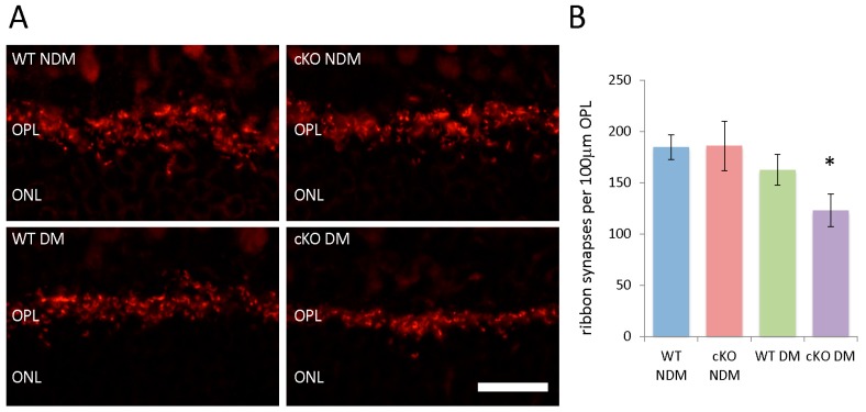
Figure 6.
(A) Retinal cryosections (20 µm) labeled with antibodies against Ribeye (red), a synaptic marker for ribbon synapses in the outer plexiform layer (OPL), in non-diabetic (NDM) and 20-week diabetic (DM) wild type (WT) and XBP1 fl/fl; Chx10-cre positive (cKO) mice; (B) Graph depicts the average number of Ribeye-positive ribbon synapses per 100 µm of the OPL for each group. There is a significant decrease in the number of ribbon synapses for XBP1 cKO DM mice. n = 3 for all groups; Mean ± S.D.; scale bar = 50 µm; * p < 0.05. ONL, outer nuclear layer.

