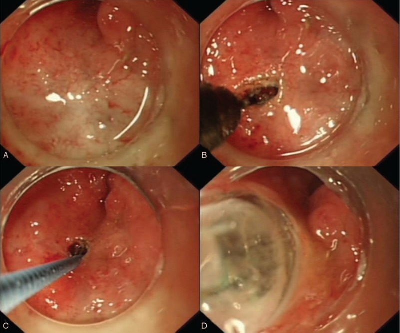
Figure 2.

(A) Endoscopic view of the expected position of the lumen. (B) A small incision was made by using a needle knife. (C) A guidewire was passed through the small incision. (D) The stenosis was sequentially dilated to a maximum diameter of 18 mm.
