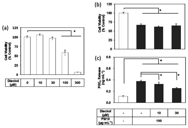
Figure 7.
Effects of dieckol on the viability and PGE2 release of HaCaT keratinocytes exposed to PM10. Cells were treated with dieckol at varied concentrations for 48 h for the viability assay (a). Cells were treated with 100 μg mL−1 PM10 in the presence or absence of dieckol at indicated concentrations for 48 h for the viability assay (b) and the PGE2 assays (c). Data are presented as a mean ± SD (n = 4). All treatments were compared to the PM10 control using one-way ANOVA followed by Dunnett’s test. * p < 0.05.

