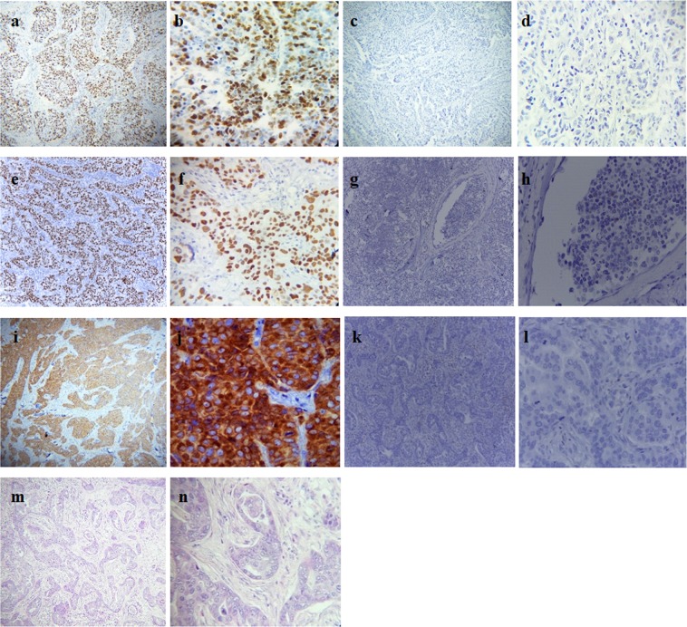
Figure 1.
Monograghs. (a and b) Nuclear positively stained for Ki-67 at 10x and 40x *hpf respectively; (c and d) Nuclear negatively stained for Ki-67 at 10x and 40x hpf respectively; (e and f) Nuclear positively stained for p53 at 10x and 40x hpf respectively; (g and h) Nuclear negatively stained for p53 at 10x and 40x hpf respectively; (i and j) Nuclear membrane positively stained for BCL-2 at 10x and 40x hpf respectively: (k and l) nuclear membrane negatively stained for BCL-2 at 10x and 40x hpf respectively; (m and n) H&E staining for infiltrating ductal carcinoma (IDC) at 10x and 40x hpf respectively. *hpf: High-power field.

