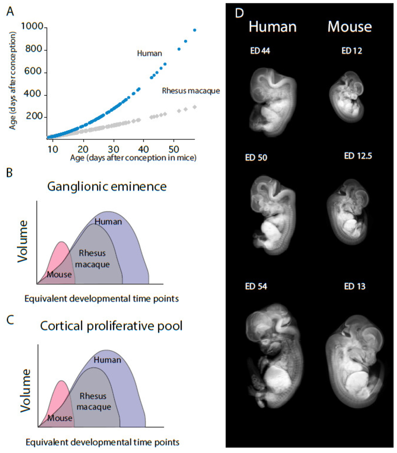
Figure 10. Extended duration of cortical and GE neurogenesis in primates versus rodents.
(A) The timing of developmental transformations (expressed as age in days after conception in macaques and humans) is regressed against equivalent developmental transformations found in mice (Workman et al., 2013; Charvet et al., 2017b, Charvet and Finlay, 2018; Charvet et al., 2018). The timing of developmental transformations is instrumental in identifying corresponding ages across species. Such an approach permits identifying variation in the timing of select developmental processes after controlling for variation in developmental schedules across species. Using this approach, Charvet et al. (2017a) found that the ganglionic eminence (GE) (B) and cortical proliferative pool (C) grow for significantly longer in humans and macaques once overall differences in the duration of developmental schedules are controlled for (see Workman et al., 2013; Charvet et al., 2017a; Charvet and Finlay, 2018; Charvet et al., 2018). (D) Examples of equivalent developmental time points are shown from reconstructed structural MRI scans and micro CT-scans. Micro CT-scans of prenatal mice are from Wong et al., 2012. Structural MR scans of prenatal humans are from the multi-dimensional human embryo project (http://embryo.soad.umich.edu/index.html).

