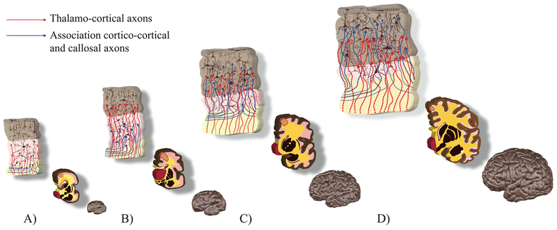
Figure 11. Summary illustration of transient fetal zones at 27 (A), 32 (B), 36 (C) and 42 (D) GW.
Upper and middle row: the cortical plate in brown, the subplate in pink, the intermediate zone in yellow. Major histogenetic events (upper row) are orange insets from the middle row: neurons in brown, thalamo-cortical axons in red, cortico-cortical and callosal axons in blue. Associated changes in cortical morphology (gyrification and surface growth) can be seen in the bottom row. Dendritic arborisation of cortical and subplate neurons is painted according to (Mrzljak et al., 1988, 1992). Development of cortical connectivity was illustrated according to the work of Kostovic and group (for reviews see (Kostovic and Jovanov-Milosevic, 2006; Kostovic and Vasung, 2009; Vasung et al., 2010)). Upper and middle rows were painted using the template coronal slices of the brains used in Vasung et al., 2016.

