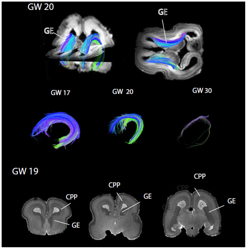
Figure 9. Diffusion MR tractography of the ganglionic eminence (GE) from gestational week (GW) 17 to 30 in humans.
The GE wanes between GW 20 and 30. Coronal planes from a structural T1w MRI scans of a human at GW19 show cell dense regions consisting of the GE and cortical proliferative pool (CPP). Comparing the growth of these two proliferative pools throughout development in humans, macaques, and mice permits identifying evolutionary changes in neurogenesis timing across species. The structural MRI scan is made available by the Allen Institute for Brain Science (Miller et al., 2014), and is available at: http://download.alleninstitute.org/brainspan/MRI_DTI_data_for_prenatal_specimens/. Image credit: Allen Institute.

