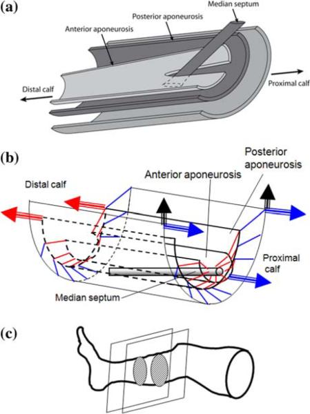
FIGURE 6.
Anatomy and function of human calf muscles. (a) Simplified schematic showing the arrangement of gastrocnemius and soleus muscle aponeuroses and the median septum. (b) Wire diagram showing individual fiber orientation (represented by short line segments; blue for gastrocnemius, red for soleus), and the direction (denoted by arrows) of forces exerted by the corresponding tendons upon muscle contraction. (c) Location of calf section studied here.

