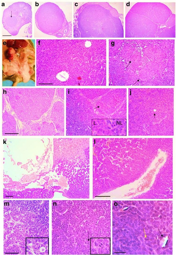
Fig. 6.
Aging APC2-deficient mice develop adult GCTs. Tumours ranged in size from (a) small in situ tumours (arrow) to (b) small tumours of normal ovarian size, (c, d) small but macroscopically visible tumours or (e) a large macroscopic tumour (arrow). The tumours displayed varying histologic patterns such as (f) follicular, (g) nodular (arrows), (h) insular, (i) luteinized (arrow: luteinized area shown at 4X original magnification in inset, L: luteinized, NL: non-luteinized), (j) diffuse (arrow: Call-Exner body), and (k) cystic patterns. The tumours were (l) highly vascularized, (m) anaplastic, (n) mitotic (black arrow), and showed (o) Call-Exner bodies (black arrow) and coffee-bean nuclei (yellow arrow). Bars a-d = 500 μm, f-l = 100 μm, m-n = 50 μm, o = 20 μm

