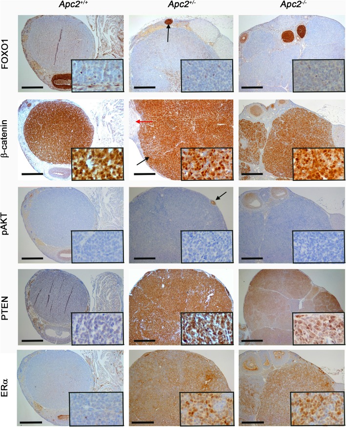
Fig. 8.
Molecular characterization (FOXO1, β-catenin, p-AKT, PTEN, ERα) of GCTs developing in aged (12–18 month Apc2 deficient ovaries. Representative photomicrographs of immunohistochemical staining of GCTs developing in Apc2+/+ ovaries (left column) and in Apc2+/− and Apc2−/− (right column) mice for FOXO1 showing weak staining in GCT, in contrast to a growing follicle (black arrow), β-catenin showing intense staining in GCT (black arrow) as compared to a corpus luteum (red arrow), p-AKT which was absent in GCT as compared to a growing follicle (black arrow), PTEN and estrogen receptor alpha (ERα). Note all genotypes also carried an Apc hypomorphic allele. Bar = 500 μm for main panel, 50 μm for insets. To maintain resolution, insets have been cropped from a representative area of a separate high power photograph, rather than a magnification of the main panel figure

