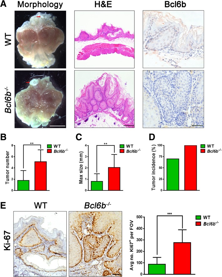
Fig. 2.
Bcl6b−/− mice developed more gastric tumours following BaP treatment. (a) Representative images showing gross morphology (Scale bar, 5 mm), H&E staining (Scale bar, 100 μm) and immunostaining of Bcl6b expression (Scale bar, 50 μm) from stomachs harbouring BaP-induced tumours (27 week) from Bcl6b−/− mice and WT controls. The gastric tumour granules were indicated by red arrows. (b-d) Tumour number (b), maximal size (c), and tumour incidence (d) in the stomachs of WT and Bcl6b−/− mice after intragastric administration of BaP (27 week). (e) Representative images and quantification of Ki-67-immunopositive cells in Bap-induced gastric tumours (27 week) from Bcl6b−/− mice and WT controls. Scale bar, 50 μm. Data are presented as the mean ± SD; n = 10 per group. **P < 0.01; ***P < 0.001; unpaired two-tailed Student’s t tests

