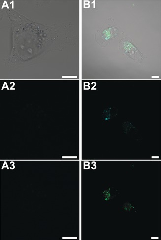
Figure 4.

Live confocal fluorescence microscopy images of MDA‐MB‐231 breast cancer cells incubated with A) fluorescein‐labeled WHW protein and B) fluorescein‐labeled WHW‐CuNCs. 1. Merged image of DIC, blue and green channel; 2. Blue channel (λexc: 405 nm); 3. Green channel (λexc: 488 nm). Scale bars: 10 μm.
