
Figure 2.
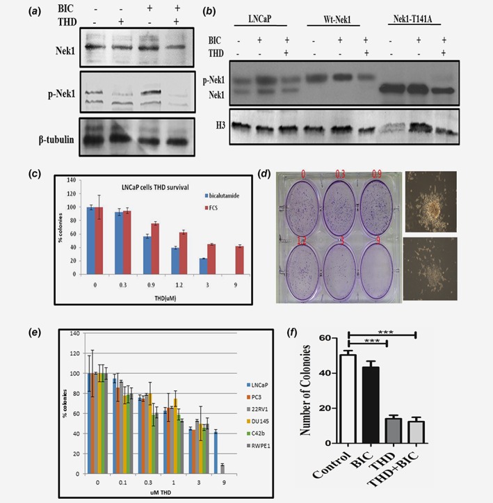
(a)–(b) Phosphorylation of Nek1 in parental LNCaP cells and overexpressing Nek1 (wt) and T141A mutant after incubation with bicalutamide +/− THD. The WB in panel b was probed with the pNek1 antiserum before affinity purification, so that it also reacts with the unphosphorylated Nek1, although more weakly. (c)–(d) AI colonies do not form with THD (concentration dependent). LNCap cells were plated in duplicates in 6‐well plates at 4000 cells with bicalutamide (10 nM) to score developing AI colonies, or 400 cells in control medium (FCS) to monitor for general clonogenic inhibition by THD. (d) An example of crystal violet‐stained plate from which the quantitation in panel C was derived. Here the AI colonies incubated with increasing concentrations of THD were scored after 3 weeks. After 1 month, a typical AI colony is shown in d (right top) without THD, or with addition of 3 μM THD (right bottom). (e) Percent of colonies treated with different conc. of THD in different PCa cell lines with increasing concentrations of THD. F. TRAMP‐C2 cells do not form AI colonies when cultured with THD. Clonogenic assays of TRAMP‐C2 cells untreated (control) or treated with bicalutamide (10 μM), THD (5 μM), or combination are displayed. [Color figure can be viewed at wileyonlinelibrary.com]