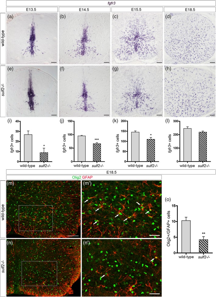
Figure 10.

Sulf2 depletion impairs early wave of AP production but does not cause major defect of astrocytogenesis. All images show transverse spinal cord sections. (a–h) Detection of parenchymal cells expressing fgfr3 mRNA in wild‐type (a–d) and sulf2−/− (e–h) embryos at E13.5 (a, e), E14.5 (b, f), E15.5 (c, g), and E18.5 (d, h). (i–l) Quantification of fgfr3+ parenchymal cells in wild‐type and sulf2−/− embryos at E13.5 (i; wild‐type n = 7; sulf2−/− n = 5), E14.5 (j; wild‐type n = 6; sulf2−/− n = 4), E15.5 (k; n = 5 for each genotype) and E18.5 (l; n = 5 for each genotype). (m–n′) Double detection of Olig2 (green) and GFAP (red) in wild‐type (m, m′) and sulf2−/− (n, n′) spinal cords at E18.5. m′ and n′ show higher magnification of the area framed in m and n, respectively. Arrows in m′ and n′ point to Olig2+/GFAP+ cells. (o) Quantification of Olig2+/GFAP+ cells in the ventral spinal cord of E18.5 wild‐type and sulf2−/− embryos (n = 5 for each genotype). Data are presented as mean ± SEM (***p < .001, **p < .01, and *p < .05). Scale bars = 100 μm in a–h, m, n and 50 μm in m′, n′ [Color figure can be viewed at wileyonlinelibrary.com]
