Figure 2.

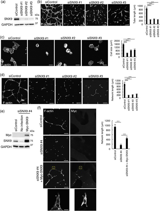
SNX9 is essential for tube formation in HUVECs. (a) Western blot analysis of HUVEC lysates 72 hr posttransfection of siRNAs. (b) Confocal images and quantitation of tube formation. HUVECs seeded on collagen I gel were treated with control, or SNX9 siRNAs (siSNX9 #1–#3) and packed on collagen I followed by VEGF stimulation for 66 hr. HUVECs were visualized by the staining for F‐actin with rhodamine‐labeled phalloidin. Bars, 200 µm. A total of 30 of the tubes from three independent experiments were analyzed. Data shows the mean ± SEM. ***p < .001. (c) Confocal images and quantitation of cell spreading. HUVECs were seeded on the Matrigel followed by incubation for 1 hr. The cells were visualized by staining of F‐actin with rhodamine‐labeled phalloidin. Bars, 20 µm. The size of 50 cells from three independent experiments were analyzed. Data shown are the mean ± SEM. ***p < .001. (d) Confocal images and quantitation of network formation. HUVECs were seeded on the Matrigel followed by incubation for 12 hr. The cells were visualized by staining for F‐actin with rhodamine‐labeled phalloidin. Bars, 200 µm. A total of 30 of networks from three independent experiments were analyzed. Data shown are the mean ± SEM. ***p < .001. (e,f) Rescue experiments for SNX9 knockdown: (e) Western blot analysis of cell lysates of HUVECs infected with siRNA resistant‐Myc‐SNX9‐carrying lentivirus. (f) Confocal images and quantitation of network formation of HUVECs infected with siRNA resistant‐Myc‐SNX9‐carrying lentivirus (f). HUVECs were seeded on the Matrigel followed by incubation for 12 hr. Cells were visualized by the staining for F‐actin with rhodamine‐labeled phalloidin. The Myc‐SNX9 was labeled with anti‐Myc antibody. Bars, 200 µm. Magnifications of the squared areas are shown in the lower panels (bars, 20 µm). A total of 30 of the tubes from three independent experiments were analyzed. Data shown are the mean ± SEM. ***p < .001. HUVEC: human umbilical vein endothelial cells; SEM: standard error of mean; siRNA: small interfering RNA; SNX9: sorting nexin 9; VEGF: vascular endothelial growth factor [Color figure can be viewed at wileyonlinelibrary.com]
