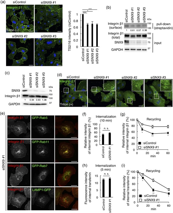
Figure 3.

SNX9 regulates localization of cell surface integrin β1 and the recycling pathway of the cargo in HUVECs. (a) Confocal images and the quantitation of HUVECs fixed after 72 hr transfection of siRNA and stained for integrin β1 by Alexa488‐conjugated TS2/16, without membrane permeabilization. Bars, 20 µm. Fifty cells from three independent experiments were analyzed. Data shown are the mean ± SEM. ***p < .001; *p < .05. (b) Surface proteins were biotinylated, collected with streptavidin beads, and the total and cell surface integrin β1 were detected by TS2/16. The numbers indicate the band intensity of surface/total integrin β1 (normalized to siControl). (c) Western blots of HUVEC lysates 72 hr posttransfection of siRNAs. The numbers indicate the band intensity of total integrin β1/GAPDH (normalized to siControl). (d) Confocal images of HUVECs fixed after 72 hr transfection of siRNA, permeabilized, and stained for integrin β1 by P5D2. Bars, 20 µm. Inset: magnifications of the squared areas. (e) Confocal images of SNX9 knockdown HUVECs expressing the GFP‐tagged organelle markers: Rab5 (early endosomes), Rab11 (recycling endosomes), Rab7 (late endosomes), and LAMP1 (lysosomes) by lentiviral infection. Bars, 20 µm. (f) Internalization of integrin β1. HUVECs were treated with control siRNA and SNX9 siRNA #1 for 72 hr. Surface integrin β1 was labeled with Alexa488‐TS2/16 and chased in medium for 10 min. The fluorescence intensity of Alexa488‐TS2/16 is shown as a percentage of that at 0 min (the fluorescence intensity on the cell surface before quenching). Fifty cells from three independent experiments were analyzed. Data shown are the mean ± SEM. n. s., not significant. (g) Recycling of integrin β1. HUVECs were treated with the control siRNA and SNX9 siRNA #1 for 72 hr. The fluorescence intensity of Alexa488‐TS2/16 is shown as a percentage of that at 0 min. Fifty cells from three independent experiments were analyzed. Data shown are the mean ± SEM. **, p < .01; *, p < .05. (h) Internalization of transferrin. The HUVECs were treated with the control siRNA and SNX9 siRNA #1 for 72 hr. The cells were incubated with Alexa488‐Tfn for 5 min, acid‐washed, and fixed. The fluorescence intensity of Alexa488‐Tfn is shown. Fifty cells from three independent experiments were analyzed. Data shown are the mean ± SEM. n. s., not significant. (i) Recycling of transferrin. The HUVECs were treated with control siRNA and SNX9 siRNA #1 for 72 hr. The fluorescence intensity of Alexa488‐Tfn is shown as the percentage of that at 0 min. Fifty cells from three independent experiments were analyzed. Data shown are the mean ± SEM. **p < .01; *p < .05. GAPDH: glyceraldehyde 3‐phosphate dehydrogenase; HUVEC: human umbilical vein endothelial cells; SEM: standard error of mean; siRNA: small interfering RNA; SNX9: sorting nexin 9 [Color figure can be viewed at wileyonlinelibrary.com]
