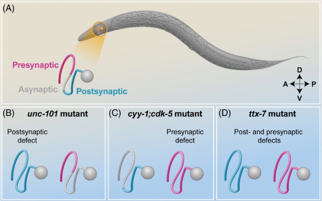
Figure 2.

Compartmentalization of synaptic molecules in the Ring interneurons A (RIAs). (A) Schematic representation of Caenorhabditis elegans in lateral view; the head is on the left, the tail is on the right, the dorsal side is up and the ventral side is down. The RIA interneurons are located in the head of the animal (orange) and the left RIA is schematically represented. The diagram of the wild‐type RIA neuron shows the synaptic compartmentalization of the RIA neurite, with the exclusively postsynaptic proximal region (blue), the asynaptic isthmus region (grey), and the distal region mixed with presynaptic and postsynaptic sites (magenta); (B) In unc-101 mutant animals the postsynaptic region, defined by the localization of the glutamate receptor GLR‐1 (blue), extends to the entire length of the neurite as a result of the lack of receptor retrieval from the distal region, whereas the distal presynaptic region (magenta), defined by the localization of the Ras‐associated binding protein RAB‐3, is unaltered; (C) Conversely, in double mutant cyy-1;cdk-5 animals the presynaptic molecules diffuse towards the dendritic compartment in a dynein‐dependent manner, leaving the distribution of postsynaptic receptors unchanged; (D) Finally, in ttx-7 mutant animals, both the distal presynaptic regions and postsynaptic regions are altered and redistributed throughout the neurite.
