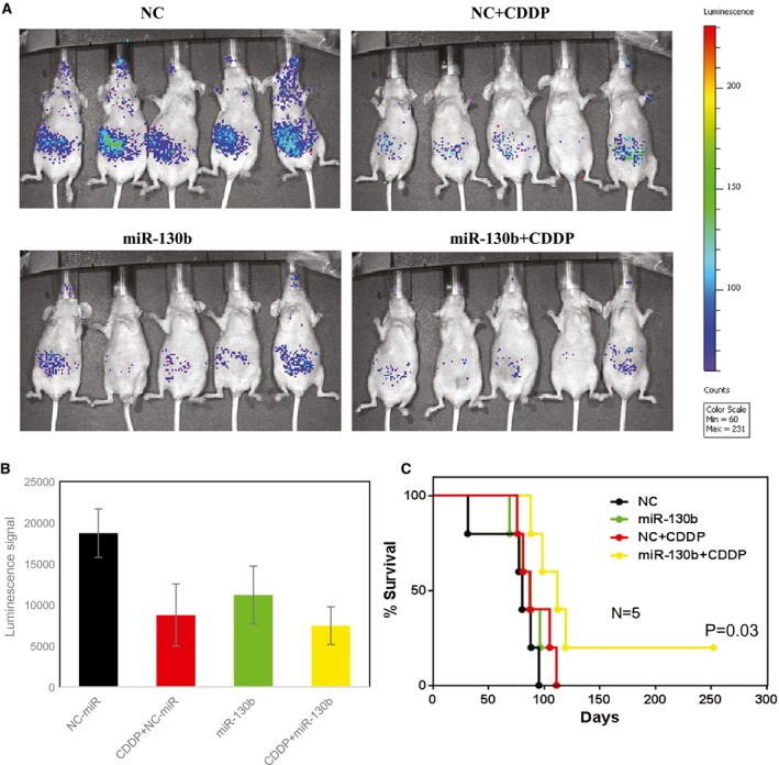
Figure 4.

In vivo chemosensitization of ovarian tumors by 1,2‐dioleoyl‐sn‐glycero‐3‐phosphatidylcholine (DOPC)–microRNA 130b (miR‐130b) is illustrated in an orthotopic mouse model of ovarian cancer. Athymic nude mice bearing OVCAR8 xenograft tumors were treated with DOPC–miR‐130b and/or cisplatin (CDDP) (miR‐130b + CDDP) twice weekly for 5 weeks. Normal control (NC) mice treated with DOPC–miR were used as controls. (A) In these images (from week 3), green corresponds to the highest signal intensity (and, thus, to tumor burden), and blue corresponds to the lowest signal intensity. Bar charts illustrate (B) the quantification of luminescence signal intensities and (C) Kaplan‐Meier curves for the chemosensitization study.
