Figure 6.

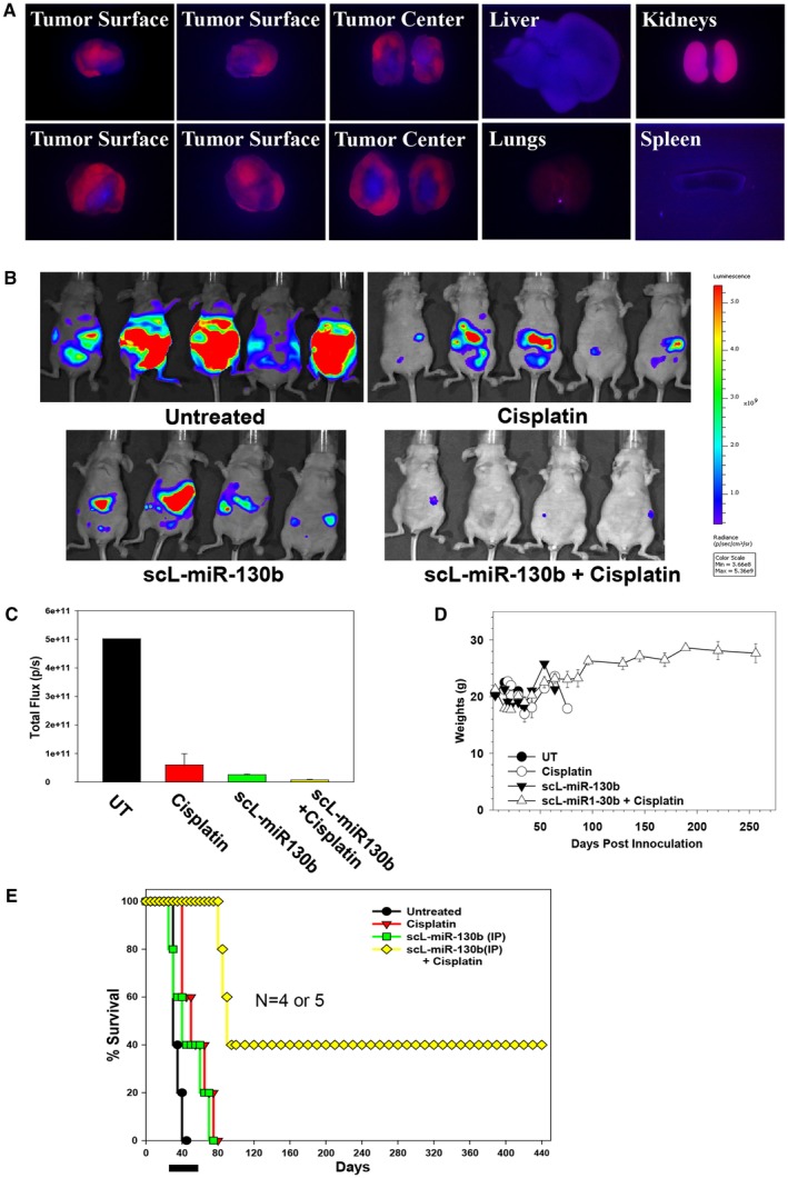
In vivo tumor targeting was achieved by systemically administered, tumor‐targeted nanocomplex (scL)‐microRNA and the chemosensitization of ovarian tumors by scL–microRNA 130b (miR‐130b) in an orthotopic mouse model of ovarian cancer. (A) Athymic nude mice bearing HEYA8 subcutaneous xenograft tumors were injected once into the tail vein with 25 μg indocarbocyanine (cy5)‐labeled, tumor‐targeted nanocomplex. Forty hours after injection, the dorsal and ventral sides and the central portion (cross‐section) of 2 individual tumors (liver, lungs, kidneys, and spleen) were imaged using an in vivo fluorescence imaging system with a cy5 detection filter. (B) Athymic nude mice bearing HEYA8 subcutaneous xenograft tumors were treated with scL–miR‐130b and/or cisplatin twice weekly for 5 weeks. Untreated animals were used as controls. The animals were imaged on day 42. Red corresponds to the highest signal intensity (maximum [max]) and, thus, tumor burden; and purple corresponds to the lowest signal intensity (minimum [min]). (C) Quantification of Xenogen signal intensities for the images were obtained 42 days post‐tumor cell inoculation. (D) The weights of the mice were monitored on a regular basis. (E) Kaplan‐Meier curves were generated from the in vivo chemosensitization study. The black bar indicates the duration of treatment. IP indicates intraperitoneal; UT, untreated.
