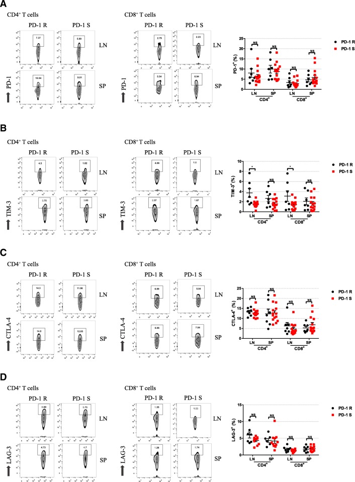
Fig. 5.
Higher frequencies of TIM-3-expressing T cells were observed in the PD-1R group. The expression of checkpoint inhibitors on CD4+ and CD8+ T cells was analyzed by flow cytometry for PD-1S group and PD-1R group. Representative flow cytometry dot plots show the analysis of checkpoint inhibitors expression on CD4+ and CD8+ T cells. The frequencies of PD-1+(a), TIM-3+(b), CTLA-4+(c) and LAG-3+(d) cells are shown. The data show that TIM-3 expression was significantly increased in the CD4+ and CD8+ cells in the LN and SP of the PD-1R group compared with PD-1 S group. All data represent the average ± SEM. Statistical significance was determined by Student’s t test, *P < 0.05, **P < 0.01

