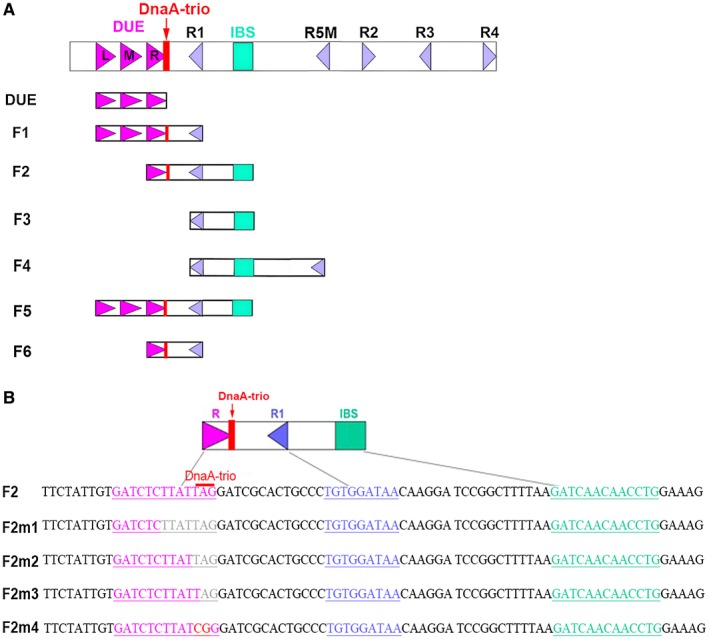
Figure 6.

Features of oriC mutations. A. Structure of oriC showing the AT‐rich 13 bp DUE regions (purple arrows), DnaA boxes (blue triangles), DnaA‐trio (red rectangle) and IBS (green rectangle). DUE, F1, F2, F3 and F4 indicate the truncated oriC constructs. B. Overview of the oriC fragments containing mutations in the replication origin element. A portion of the DUE‐R 13 bp fragment is indicated in pink. DnaA box R1 is indicated in blue, and the IBS is indicated in green. Mutation sites are indicated in red, and deletion sites are indicated in light gray. The DnaA‐trio is indicated by a red line. [Colour figure can be viewed at wileyonlinelibrary.com]
