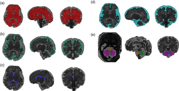
Figure 2.

Segmentation of the brain from a fetus with Down syndrome at 33+2 gestational weeks. Semi‐automated segmentation of T2‐weighted volumetric magnetic resonance images showing (a) whole brain; excluding cerebellum (red), (b) cortex (green), (c) lateral ventricles (dark blue), (d) extra cerebral cerebrospinal fluid (light blue), (e) cerebellar hemispheres (purple), cerebellar vermis (bright green), pons (yellow), and fourth ventricle (blue).97 [Colour figure can be viewed at wileyonlinelibrary.com]
