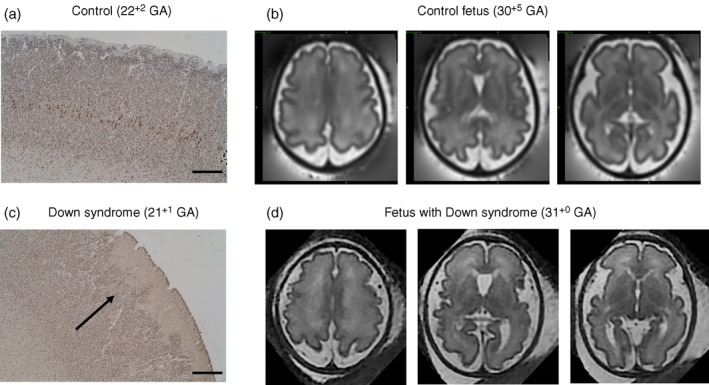
Figure 7.

Neuronal staining in the cortex of human fetal postmortem tissue. HuC/HuD, a marker for all neurons in brain from control fetus at 22+2 GA (a) and fetus with Down syndrome at 21+1 GA (c). In the fetal brain with Down syndrome (c,d), the black arrow indicates evidence of aberrant cortical folding, a ‘wavy’ pattern which is in contrast to the control brain (a,b) (Research Ethics Committee UK: 07/H0707/139). Scale bar=500μm. T2‐weighted fetal magnetic resonance imaging in the axial plane show decreased cortical folding in a fetus with Down syndrome (d), compared to an aged matched control (b). GA, gestational age expressed as weeks+days. [Colour figure can be viewed at wileyonlinelibrary.com]
