
Figure 1.
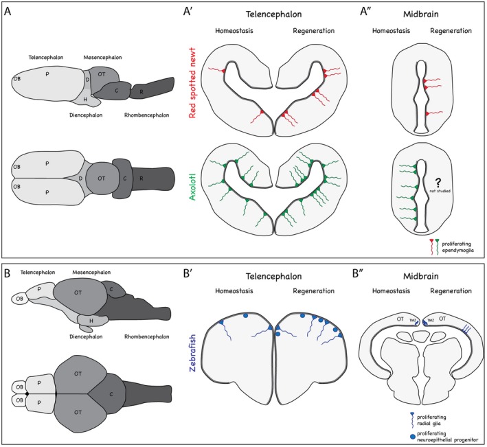
Brain structure and neurogenic niches during homeostasis and regeneration in salamanders and teleost fish. (A and B) Schematic illustrations of the lateral and dorsal views of salamander (A) and zebrafish (B) brains. OB = olfactory bulb, T = telencephalon, OT = optic tectum, D = diencephalon, H = hypothalamus, C = cerebellum, R = rhombencephalon. (A′ – B″) Cross sections through telencephalon and midbrain of red spotted newt, axolotl (A′, A″), and zebrafish (B′, B″). The left hemisphere depicts the proliferative behavior of cells during homeostasis, while the right hemisphere depicts the proliferative behavior of cells following injury. Thick grey lines indicate the ventricular zones, harboring radial glia or ependymoglia. Note the inverted organization of the teleost fish telencephalon, in which radial glia and non‐epithelial progenitors line the outside and neurons are located on the inside. (A′) In red spotted newts, ependymoglia (red) proliferate during homeostasis in confined hot spots, while in axolotl ependymoglia proliferation (green) is observed along the entire ventricular zone. In red spotted newts, ependymoglia cells at hot spots increase their proliferation rate in response to injury and additional hot spots of proliferation are generated. In axolotl, ependymoglia proliferation is increased. (A″) The midbrain of red spotted newts is quiescent during homeostasis, while axolotl midbrain ependymoglia are proliferative. Upon injury in red spotted newts, ependymoglia re‐enter the cell cycle locally where neurons were lost. Midbrain regeneration in axolotl has not been studied. (B′) In zebrafish, radial glia as well as non‐epithelial progenitors (blue) actively divide during homeostasis. Upon injury, additional radial glia and non‐epithelial progenitors are activated to proliferate. (B″) During homeostasis in the optic tectum, radial glia lining the roof of the tectal ventricle are quiescent, while neuroepithelial‐like progenitors located at the tectal marginal zone (TMZ) are proliferative. Upon injury, neuroepithelial‐like progenitors increase their proliferation and radial glia enter the cell cycle