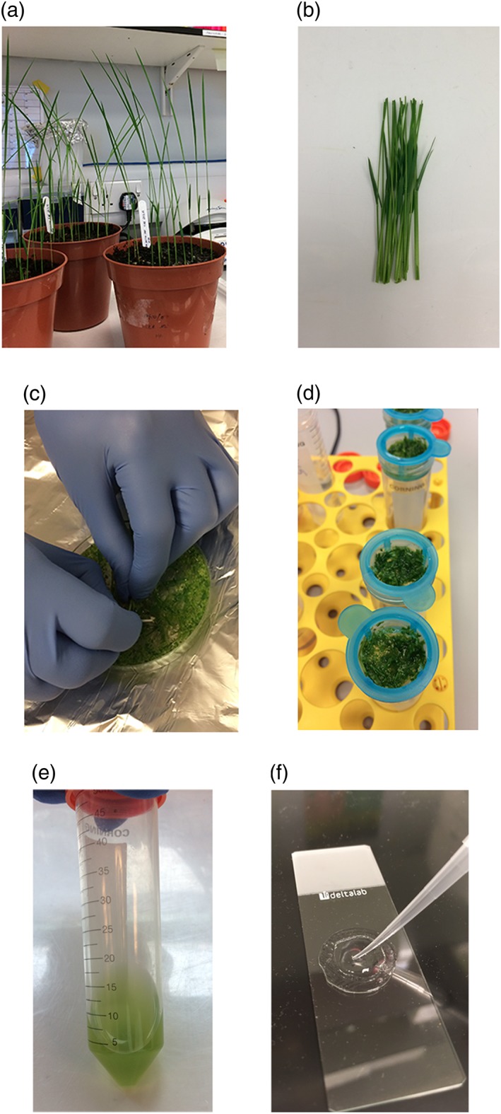
Figure 1.

Photographs of various steps in the protocol. Rice seedlings 7 days after germination (a), approximately 100 mm of rice stem and sheath tissue (b), cutting tissue into mannitol to initiate plasmolysis (c), filtration of digested tissue through 40 μm mesh (d), protoplast suspension after isolation (e), and mounting transformed protoplasts onto slides in a well constructed from observation gel (f). Scale bars represent 45 mm (a), 20 mm (b), 25 mm (c), 15 mm (d), 15 mm (e), 10 mm (f)
