Figure 2.

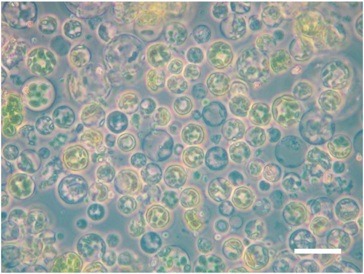
Isolated protoplasts imaged using light microscopy. Isolated protoplasts were imaged with a Zeiss Axioskop 40 microscope under 40x magnification. Scale bar represents 40 μm

Isolated protoplasts imaged using light microscopy. Isolated protoplasts were imaged with a Zeiss Axioskop 40 microscope under 40x magnification. Scale bar represents 40 μm