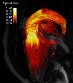
Figure 2.

Pathlines covering complete heart cycle from spiral 4D flow acquisition of patient 3, 30‐year‐old male with repaired congenital heart disease (Senning repair for transposition of great arteries), and moderately dilated right (systemic) ventricle with mild systolic dysfunction. Pathlines were released backward and forward from segmentation of right ventricle at end diastole. Three‐chamber balanced steady‐state free‐precession image is shown for orientation. Pathlines are color‐coded according to speed.
