
Figure 4.
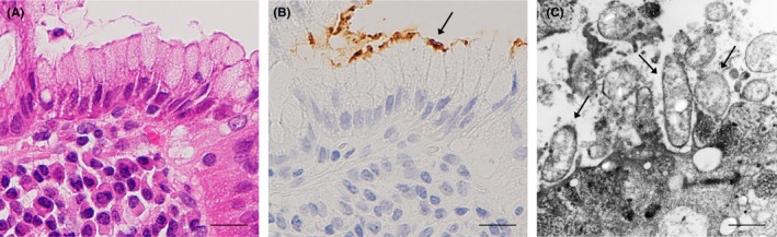
Localization of Helicobacter pylori cells in mucous layer attached to gastric foveolar epithelial cells. Serial histologic sections of a sample with prominent H. pylori infection were used for hematoxylin & eosin staining (A) and IHC with TMDU‐mAb (B) followed by immuno‐electron microscopy (C). (A and B) show an identical area of the H. pylori‐infected gastric mucosa. The area with immunoreactive H. pylori indicated by an arrow in b was subjected to immuno‐electron microscopy. (c) Several H. pylori organisms with a dense rim‐staining pattern indicated by arrows were observed on the foveolar epithelial cell. Bars: 20 µm (A and B), 1.0 µm (C)